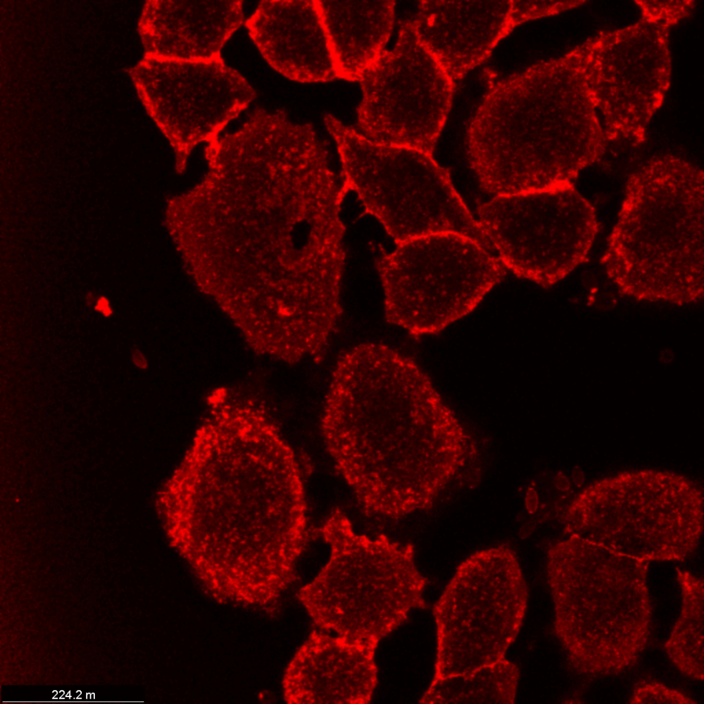
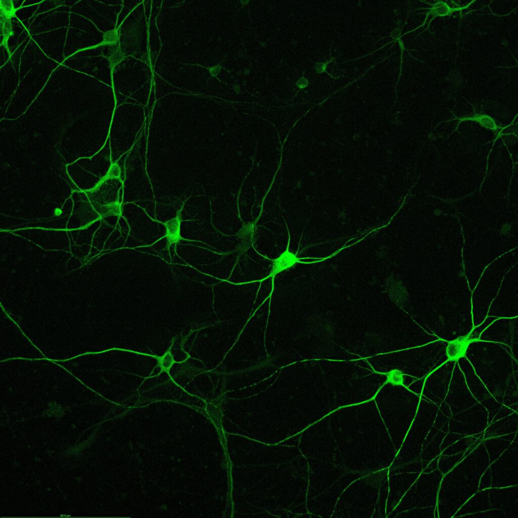
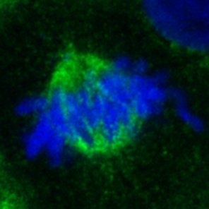
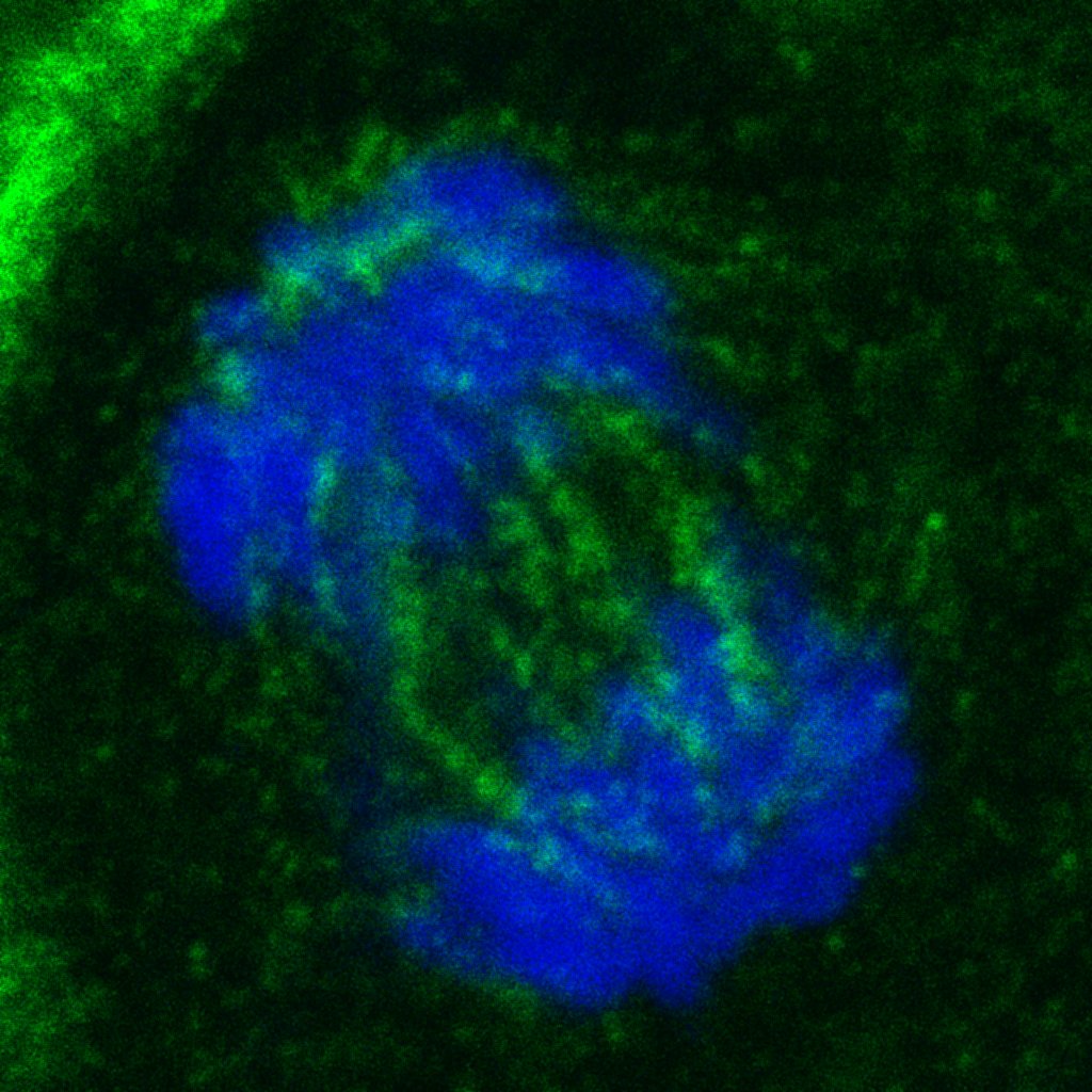
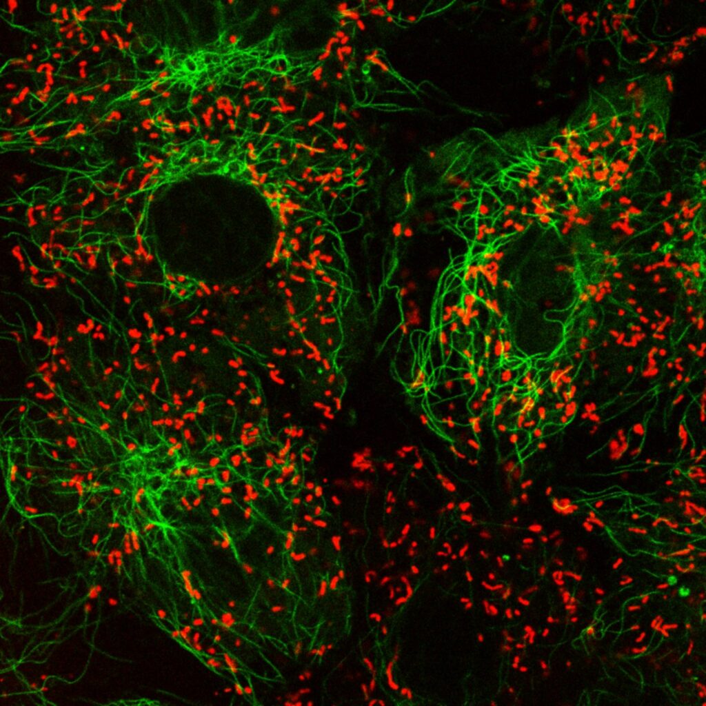
The confocal microscope is used for imaging of specific parts of the cells, such as mitochondria, tubulin network or immunofluorescent labelled proteins. The white excitation laser allows the use of a wider range of fluorescent labels and multiplexed staining; the 405 nm laser adds the possibility of using a fourth label for the nucleus and also photoconversion of specific samples. The quality of the images obtained is greatly improved by gating. The pulsed laser and hybrid detectors remove a large part of the non-specific light (reflection, scattering). The resonance scanner allows the measurement of fast events like local calcium signals using calcium indicators. Other speed-intensive techniques can also be performed.

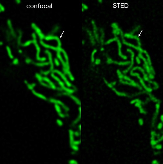
This microscope has also the possibility of super-resolution confocal imaging (STED). The two depletion lasers (592 and 775 nm) can be used to achieve better than optical resolution (~100nm).
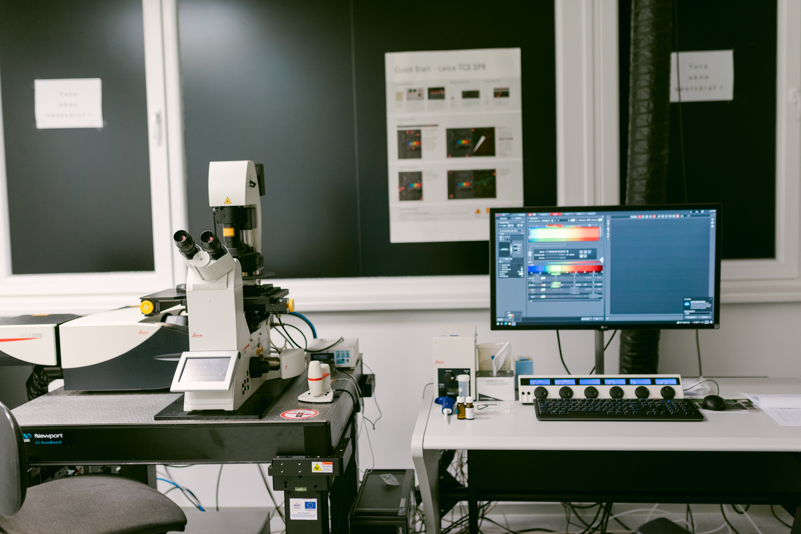
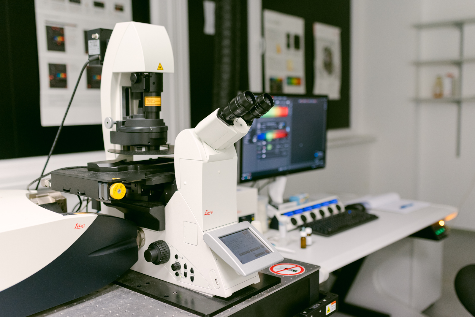
Leica TCM SP8 STED 3X Microscope STED Configuration:
- DMi8 inverted microscope
- AOBS
- Standard objectives 63x water and oil, special objective STED WHITE 100x
- Tandem scanner 8 kHz
- 2x HyD, 2x PMT, 1x TLD setectors
- Gated STED , 3D STED
- SuperZ galvo for fast 3D imaging
- Laser 405 nm
- Pulsed white laser WLL2 – 470 – 670 nm
- Laser depletion for STED: 592 nm, 660 nm, pulsed laser 775 nm
Contact: Mgr. Zuzana Ševčíková Tomášková, PhD.
Support: ITMS 26230120006